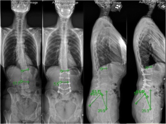
Fig. 2.

Preoperative and postoperative 36-in.-long cassette radiographs obtained in a patient who underwent a minimally invasive correction of ASD including L1/2, L2/3, L3/4, and L4/5 lateral lumbar interbody fusion and posterior L1–L5 percutaneous pedicle screws. The coronal Cobb angle reduced from 17.7° to 5.5° and lumbar lordosis increased from 49° to 57.7°
