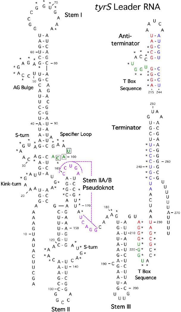
Figure 1.
Structural model of the B. subtilis tyrS leader RNA. The sequence is shown from the transcription initiation site through the termination site. The terminator form is shown, with the antiterminator form above the terminator helix. Structural domains and conserved sequence elements are labeled. Highly conserved residues are marked with asterisks. The pseudoknot pairing is shown in purple. Sequences on the 5’ side of the terminator (blue) pair with a portion of the T box sequence (red) to form the antiterminator. The residues that interact with tRNATyr (UAC Specifier Sequence and UGGU in antiterminator bulge) are shown in green. Modified from [5].

