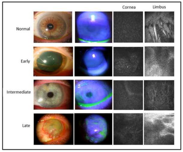
Figure 1.
Representative slit lamp photos (left column) and fluorescein staining patterns (middle left column) of normal controls and of patients with early, intermediate, and late stage limbal stem cell deficiency are shown. Representative in vivo confocal images of the corneal (middle right column) and limbal (right column) basal epithelium in normal controls and in patients with limbal stem cell deficiency are shown.

