Figure 1.
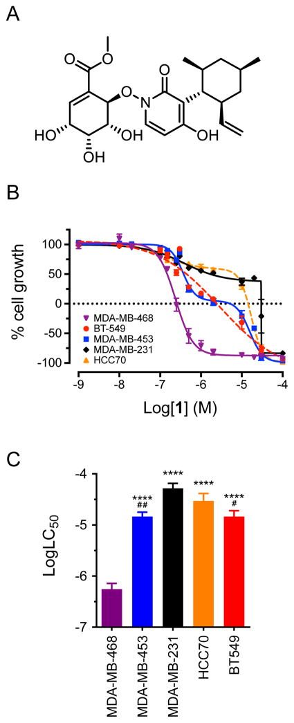
Compound 1 is selectively cytotoxic to basal-like 1 MDA-MB-468 cells. (A) Structure of 1. (B) Concentration-response curves for antiproliferative and cytotoxic effects of compound 1 (maximiscin) in five triple-negative breast cancer cell lines after 48 h treatment. Results represent mean ± SEM for at least 3 independent experiments with all concentrations tested in triplicate. (C) Comparison of log LC50 values for compound 1 in five triple-negative breast cancer cell lines. ****p < 0.0001 compared to MDA-MB-468; #p < 0.05, ##p < 0.01 compared to MDA-MB-231; one-way ANOVA with Tukey’s posthoc test. Results represent mean ± SEM for at least 3 independent experiments, each conducted with all concentrations tested in triplicate.

