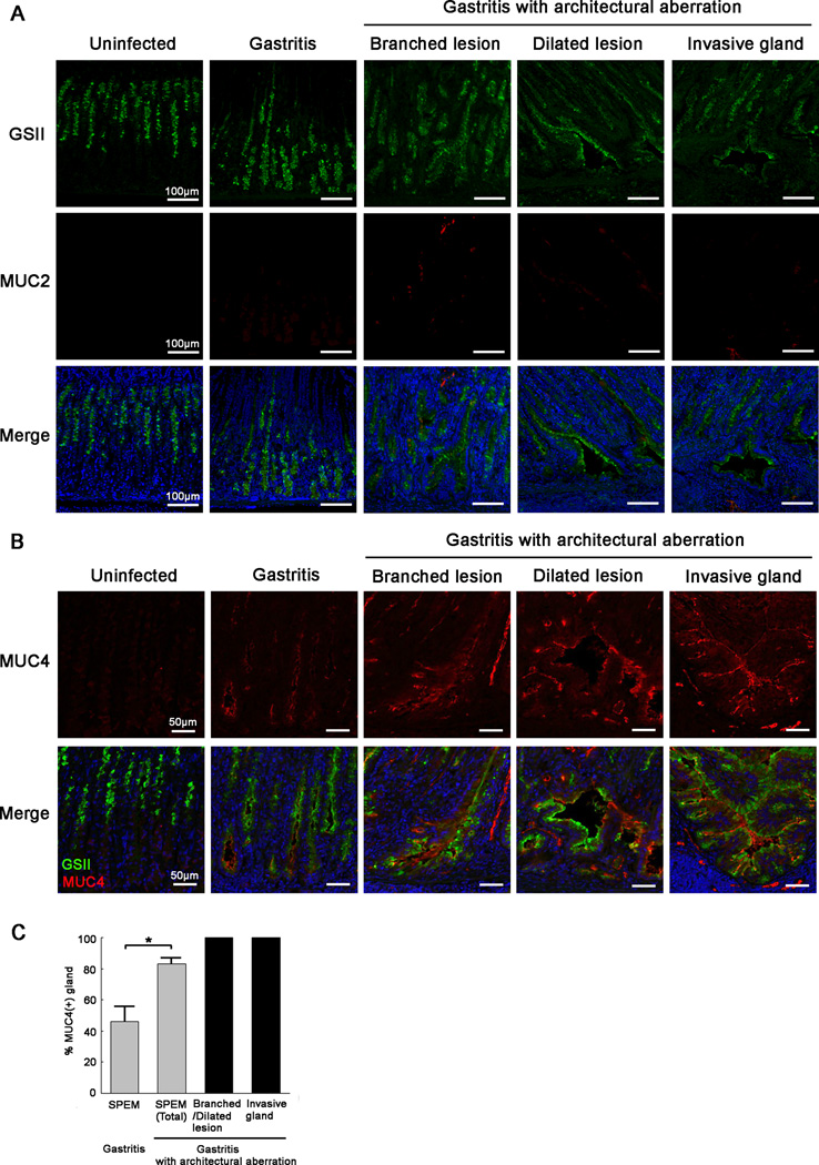
Figure 2. Progression of metaplastic lesions in gerbil gastric corpus mucosa following H. pylori 7.13 infection.
(A) Immunofluorescence staining of metaplastic glands with GSII lectin (green) and antibodies against MUC2 (red). All sections were from the corpus or intermediate zone of gerbils. Gastritis sections shown, containing glands without architectural aberration, were from representative gerbils at 6 wk post infection. Gastritis sections showing glands with architectural aberration, including branched lesions, dilated lesions and invasive glands were from representative gerbils in the corpus mucosa of a representative animal at 8 wk post infection. In uninfected gerbils, GSII expression was seen in mucous neck cells. GSII was expressed not only in cells at the base of gastritis glands without architectural aberration, but also in branched lesions, dilated lesions and invasive glands of gastritis glands with architectural aberration. MUC2 was not expressed in the normal corpus or in the corpus of animals with gastritis. Bar=100 µm.
(B) Immunofluorescence staining of SPEM with antibodies against MUC4 (red) and GSII lectin (green). All sections were from the corpus or intermediate zone of gerbils. Representative gastritis sections without architectural aberration are shown from gerbils in the corpus mucosa of a representative animal at 6 wk post infection. Branched lesions and dilated lesions were analysed from in the corpus mucosa of a representative gerbil at 12 wk post infection. Invasive glands were examined in the gastric corpus mucosa of a representative gerbil at 16 wk post infection. In uninfected gerbils, MUC4 expression was not observed. In gastritis without architectural aberration, MUC4 was weakly expressed in the sub-apical region of cells at the base of SPEM glands. In gastritis with architectural aberration, MUC4 was strongly expressed in progressive phenotypes of SPEM, including dilated lesions, branched lesions and invasive glands. Bar=50 µm.
(C) Quantitation of the percentage of MUC4-positive glands in SPEM. All gastric corpus glands were analysed in each section of all samples used in this study. SPEM was detected by GSII staining. In the presence of more severe inflammation, the percentage of MUC4-positive glands increased significantly. All glands with architectural aberration expressed MUC4 expression. All values are shown as mean±SEM (*p<0.05 vs gastritis without architectural aberration.).

