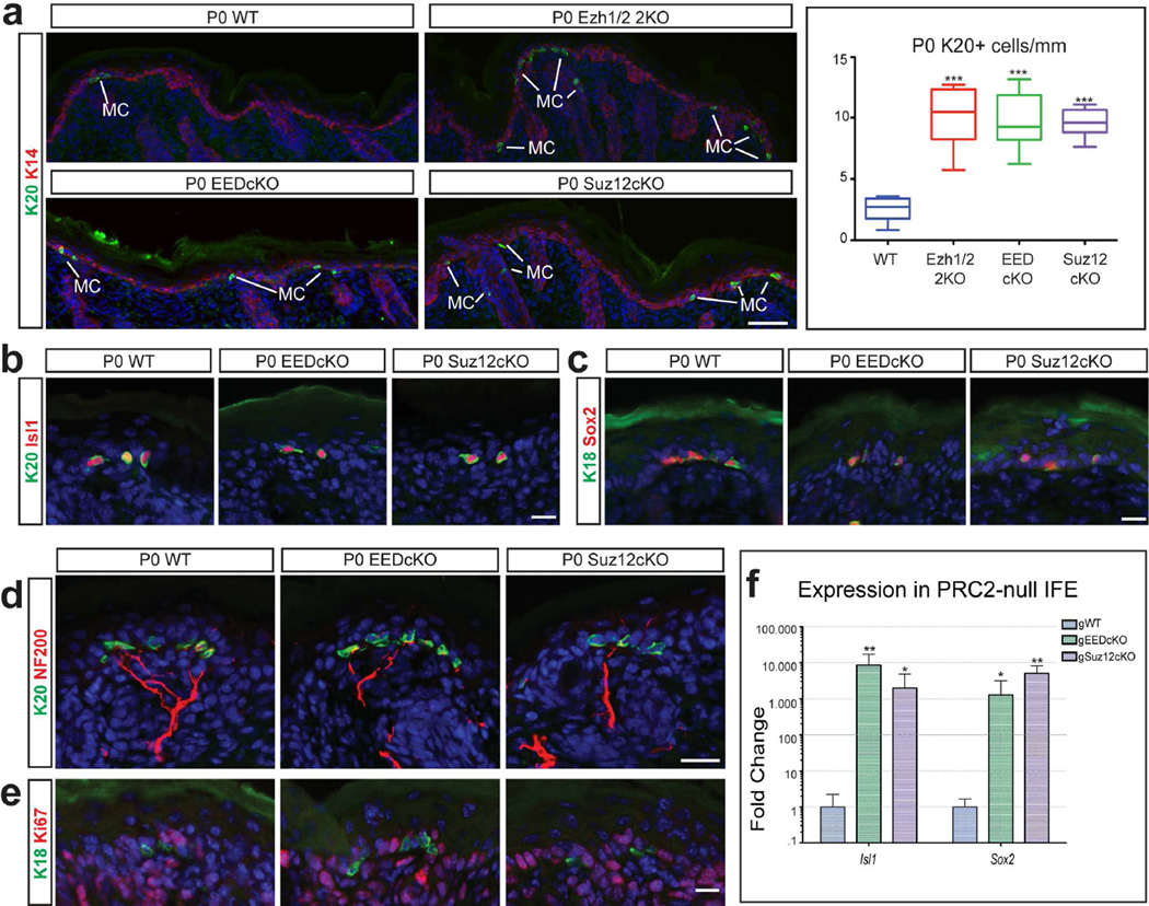
Figure 2. Loss of PRC2 results in Merkel cell expansion.
(a) Immunofluorescence for Krt20 (K20) showing a significant increase in the number of Merkel cells (MC) in P0 PRC2-null epidermis, compared to WT; quantification of number of Merkel cells (n≥3; p<0.0001). (b,c) Immunofluorescence for Merkel cell markers Krt20 (b), Isl1 (b), Krt18 (K18) (c), and Sox2 (c) showing co-expression of markers in PRC2-null Merkel cells. (d) Immunofluorescence for Krt20 and NF200 showing that the PRC2-null Merkel cells are innervated. (e) Ki67 staining showing that Merkel cells are not proliferating in P0 PRC2-null epidermis. (f) RT-qPCR in FACS-purified interfollicular epidermis (IFE) cells showing upregulation of Merkel genes Isl1 and Sox2 in P14 gWT and gPRC-null skin; (mean +/−SD; n=3; all significant, p<0.05). Scale bars: (a): 100µm: (b–e): 25µm.

