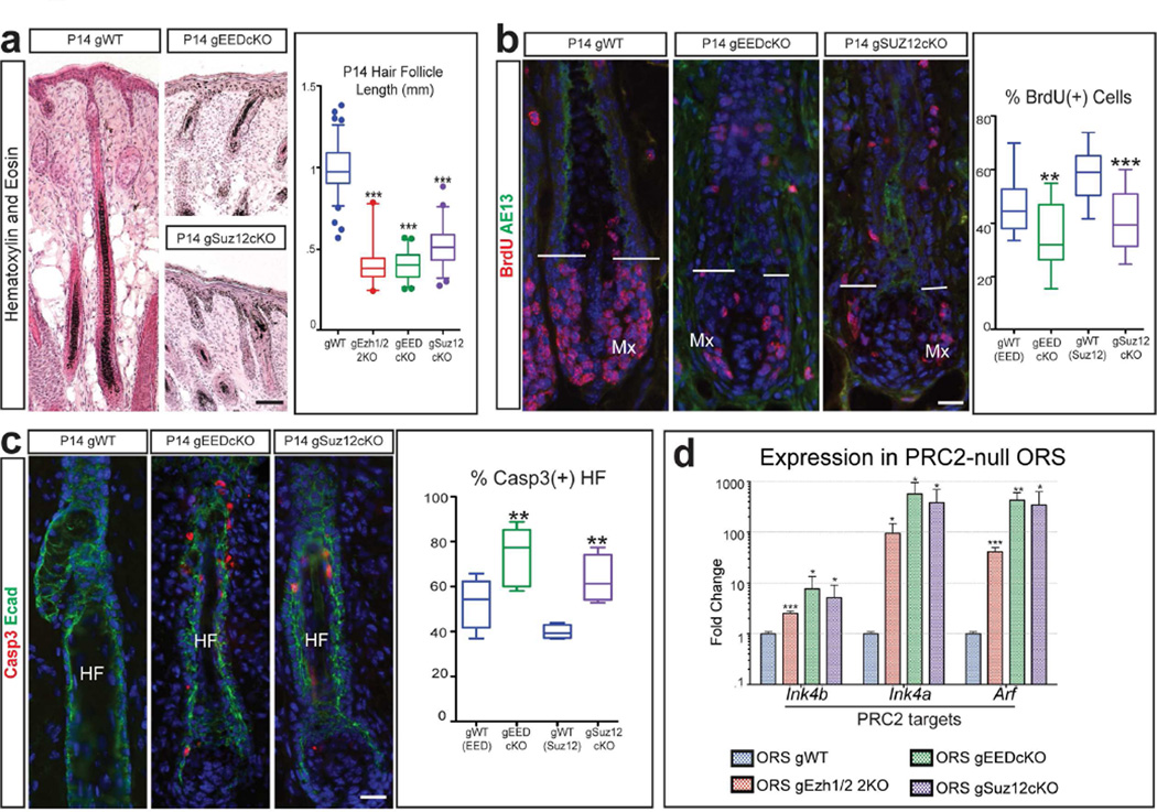
Figure 3. Loss of PRC2 causes reduced proliferation and increased apoptosis in hair follicles.
(a) Hematoxylin and Eosin showing that P14 gPRC2-null mice have shorter hair follicles (HF) than gWT; quantification of HF length (n≥2; p<0.0001). (b) BrdU staining showing reduced proliferation in the matrix (Mx) of P14 gPRC2-null HFs; quantification of percentage of BrdU(+) matrix cells (n=3; gEEDcKO, p=0.0083; gSuz12cKO, p<0.0001). AE13 marks matrix limit. (c) Activated Caspase 3 (Casp3) staining showing increased apoptosis in P14 gPRC2-null HFs, labelled with E-Cadherin (Ecad); quantification of percentage of Casp3(+) HFs (n≥2; gEEDcKO, p=0.0055; gSuz12cKO, p=0.0055). (d) RT-qPCR of FACS-purified outer root sheath (ORS) cells showing p15(INK4B), p16(INK4A), and p19(ARF) upregulation in P14 gPRC2-null mice (mean +/− SD; n=3; all significant, p<0.05). Scale bars: (a): 100µm; (b,c): 25µm.

