Fig. 8.

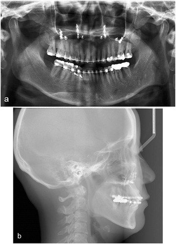
Postoperative panoramic view showing increased vertical dimension (a) and cephalometirc X-ray of case 2 showing increased interarch space of right posterior maxillary area (b)

Postoperative panoramic view showing increased vertical dimension (a) and cephalometirc X-ray of case 2 showing increased interarch space of right posterior maxillary area (b)