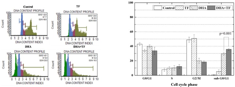
Fig. 2. Effects of dihydroartemisinin (DHA) and holotransferrin (TF) on the cell cycle of C6 cells.
Analysis of cell cycle assay with PI labeling was accomplished to quantify the percentage of C6 cells by phases. DNA fragmentation in apoptotic cells translates into the fluorescence intensity in sub-G0/G1 phase, and it is lower than that of G0/G1 cells. P-value by t-test of DHA and DHA+TF.

