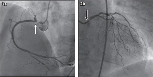
Fig. 2.

Coronary angiograms show (a) 90% stenosis in the ostium of the right coronary artery (white arrow) in the left anterior oblique view; and (b) 90% stenosis in the ostium of the left main coronary artery (black arrow) with diffuse disease in the left anterior descending and left circumflex artery in the right anterior oblique cranial view.
