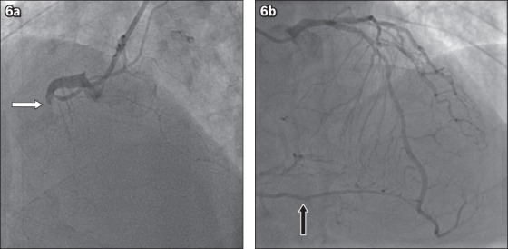
Fig. 6.

Coronary angiogram shows (a) 100% occlusion of the proximal right coronary artery (white arrow) in the left anterior oblique cranial view; and (b) collateral from the left coronary arteries (black arrow) in the left anterior oblique cranial view.
