
Fig. 1.
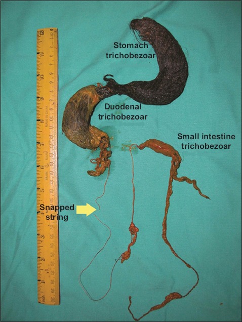
Photograph shows a large stomach trichobezoar; large duodenal trichobezoar communicating with an embedded, snapped string; and a small intestinal trichobezoar that were completely removed from the patient.
Official websites use .gov
A
.gov website belongs to an official
government organization in the United States.
Secure .gov websites use HTTPS
A lock (
) or https:// means you've safely
connected to the .gov website. Share sensitive
information only on official, secure websites.
Photograph shows a large stomach trichobezoar; large duodenal trichobezoar communicating with an embedded, snapped string; and a small intestinal trichobezoar that were completely removed from the patient.