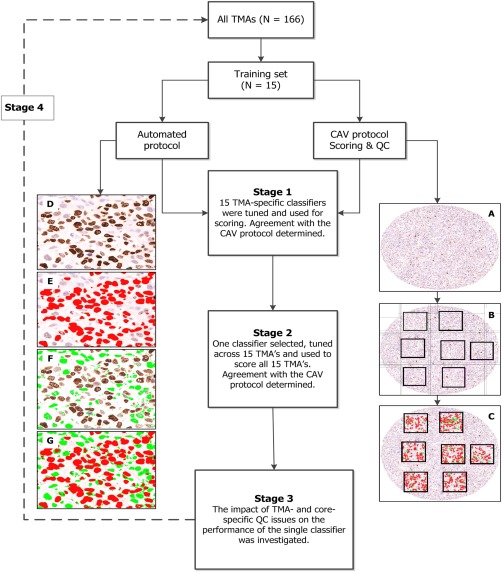
Figure 2.

Schematic representation of the stages involved in the development of a centralised scoring protocol. Of the 166 TMAs, 15 were randomly selected as the training set. Two protocols were developed and adopted for scoring: A computer‐assisted visual (CAV) and automated scoring protocols. Using the CAV protocol, a grid was used to demarcate each core and at least six well‐delineated areas of the core were counted for positive and negative nuclei (right hand panel (A) tumour core; (B) demarcation into regions by a grid and (C) counting of positive and negative nuclei within the squares) and the average score obtained. For the automated scoring protocol (Stage 1), 15 TMA‐specific classifiers were tuned (left hand panel (D) region of interest, (E) colour detection of DAB/positive nuclei, (F) colour detection of haematoxylin/negative nuclei and (G) combined detection of positive and negative nuclei) and used for scoring. In the next stage (Stage 2) one classifier was selected, tuned further, and used to score all 15 TMAs. Agreement with the CAV protocol was further tested and the impact of quality control on the performance of this classifier was then assessed (Stage 3). In the final stage (Stage 4), this classifier was applied to the scoring of all 166 TMAs in this study.
