Figure 2.

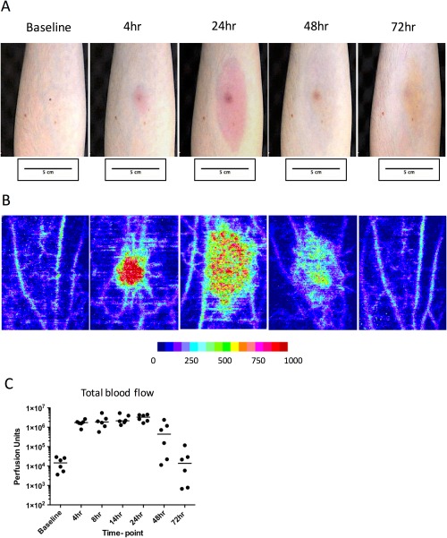
Assessment of vascular response at the site of UVKEc‐triggered resolving acute inflammation. Acute inflammation was triggered in the ventral aspect of forearm of healthy volunteers by the intradermal injection of 1.5 x 10⁷ UV‐killed E. coli (UVKEc) suspended in 100 μl of sterile saline. Vascular response at the site was assessed by laser Doppler imager (moorLDI‐HIR) which captured the camera images (A) and also generated the flux images (B). Representative flux images at baseline, 4, 24, 48 and 72 h are shown here. Flux images were analysed by moorLDI software to quantify total blood flow. Total blood flow, measured in perfusion units, is shown in panel C. Data are expressed as individual values with median; n = 6 at each time point.
