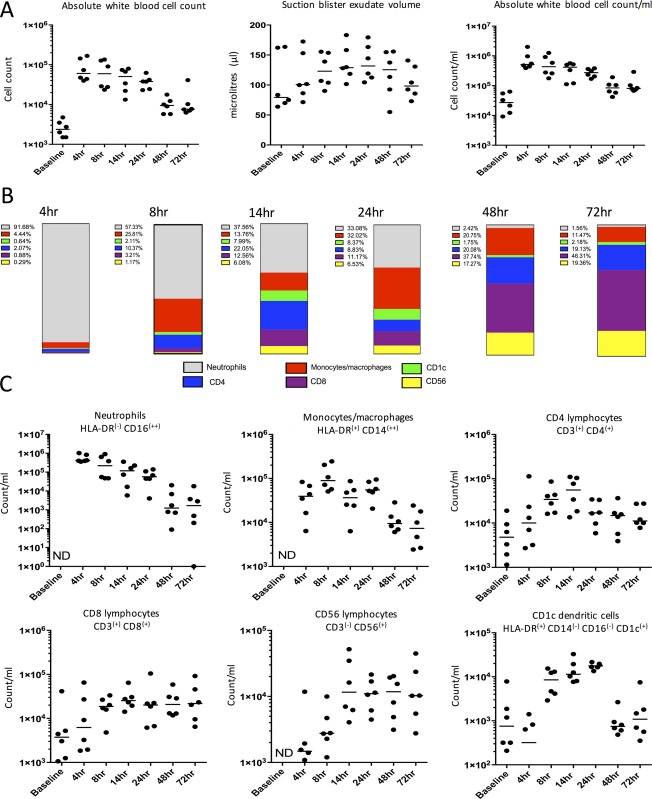
Figure 3.

Cellular profile at site of UVKEc‐triggered resolving acute inflammation. A suction blister was raised over the inflamed site by negative pressure to collect the inflammatory exudate. Exudate was centrifuged to separate cells from the supernatant containing soluble mediators. Panel A shows total white blood cells, counted manually by haemocytometer, suction blister exudate volume and total cell count per ml of exudate acquired. Panel B shows the relative proportion of neutrophils, monocytes/macrophages, CD4+ and CD8+ T lymphocytes, CD56+ NK cells and CD1c+ dendritic cells calculated at each time point, using the gating strategy illustrated in Figure 4. Panel C shows cell count/ml of the above cell populations at different time points. Data are expressed as individual values with median; n = 6 at each time point. ND = not detectable.
