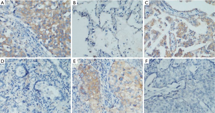
Figure 2.
Immunohistochemical analyses of C9orf86 staining. As the image above, C9orf86 was showed cytoplasm staining in lung cancer tissue (A) and no staining in matched adjacent non-cancer tissues (B), original magnification 200×; C9orf86 positive expressed (C) and negative expressed (D) in different lung adenocarcinoma tissues, original magnification 200×; C9orf86 positive expressed (E) and negative expressed (F) in different lung squamous cell carcinoma tissues, original magnification, 200×. C9orf86, chromosome 9 open reading frame 86.

