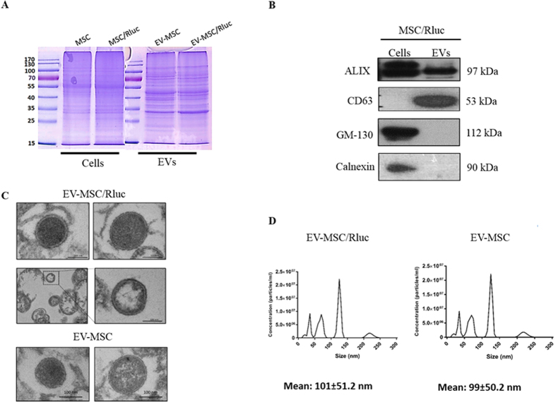
Figure 1. Characterization of EV-MSC and 1 EV-MSC/Rluc.
(A) Coomassie Brilliant Blue staining. Protein was isolated from naïve MSC and MSC/Rluc cells and their EVs. Equivalent amounts (50 μg) of protein were run on a 10% SDS gel and stained with Coomassie blue. (B) Western blotting analysis of EV marker proteins. ALIX and CD63, positive EV marker proteins, were detected in the MSC/Ruc EV. GM-130 (golgi marker) and calnexin (endoplasmic reticulum marker), negative marker proteins for EV, was not detected in MSC/Rluc EVs. (C) Analysis of EV-MSC and EV-MSC/Rluc by TEM. Image shows EVs with lipid-bilayer (Scale bars, 100 nm), (D) EV-MSC and EV-MSC/Rluc size analyzed by NanoSight. Data are expressed as the mean ± standard deviation (SD) of three independent experiments.

