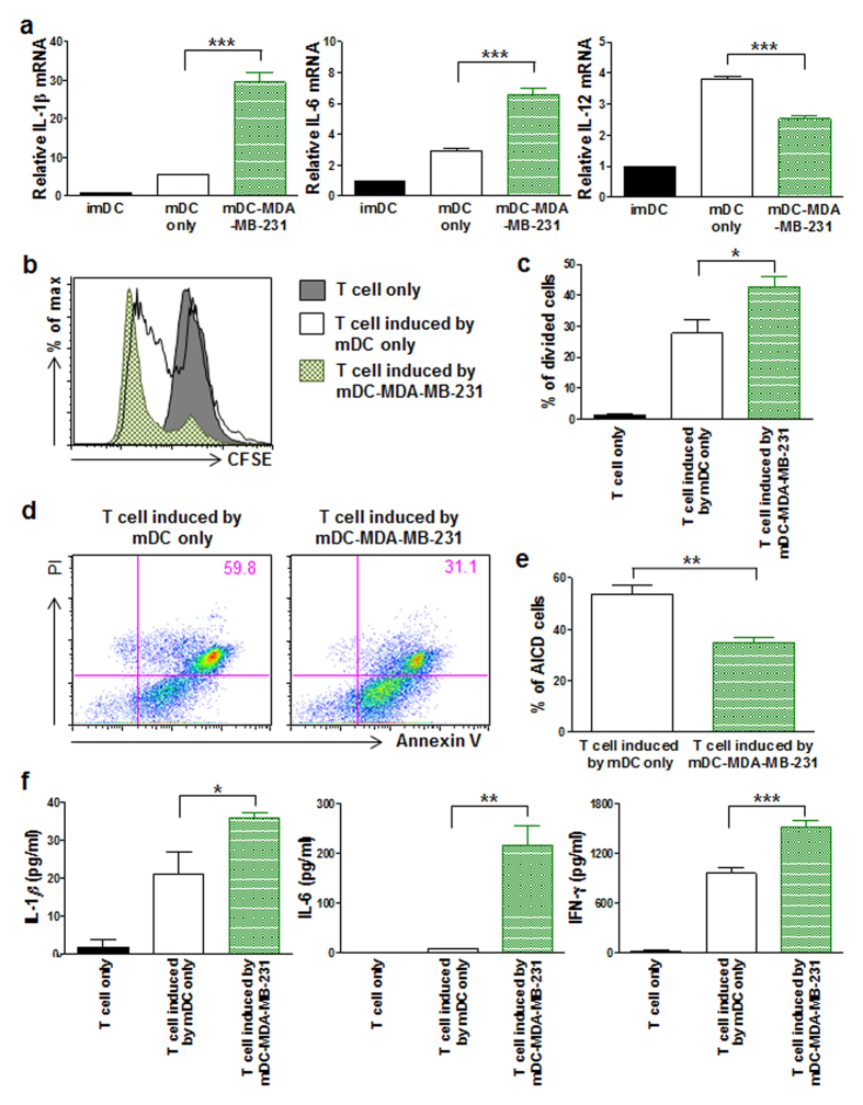
Figure 5. mDC-MDA-MB-231 expressing pro-inflammatory cytokines induces T cells to more proliferate and resistant to activation-induced cell death.
(a) The relative mRNA expressions of IL-1β, IL-6, and IL-12 in imDC, mDC only or mDC-MDA-MB-231. (b) Flow cytometric analysis of carboxyfluorescein succinimidyl ester (CFSE)-labeled CD3+ T cell only and CFSE-labeled CD3+ T cells co-cultured either with mDC only or mDC-MDA-MB-231. (c) Data represent mean ± s.e.m. of three independent experiments described in (b). (d) Flow cytometric analysis of propidium iodide (PI) and annexin V staining of CD3+ T cells activated by CD3 and CD28 antibodies in the presence of IL-2; these cells had previously been co-cultured either with mDC only or mDC-MDA-MB-231. (e) Data represent mean ± s.e.m. of three independent experiments described in (d). (f) The cytokine levels of IL-1β, IL-6 and IFN-γ secreted from CD3+ T cells only and CD3+ T cells co-cultured either with mDC only or mDC-MDA-MB-231. Data represent mean ± s.e.m. of three independent experiments (a,f). *P < 0.05, **P < 0.01 and ***P < 0.001.

