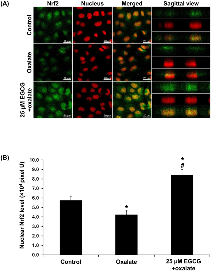
Figure 7. EGCG induced Nrf2 activation to prevent oxalate-induced EMT.
(A) The cells were stained by indirect immunofluorescence assay using a specific antibody against Nrf2 (shown in green), whereas nuclei were counterstained with propidium iodide (shown in red). The images were captured under a laser-scanning confocal microscope. In merged fields and sagittal views, nuclear localization/translocation of Nrf2 is shown in yellow (in the nuclei). Original magnification power = 630X. (B) Fluorescence intensity representing Nrf2 level was measured and analyzed from 10 random high-power fields (HPF) and at least 100 cells in each sample. N = 3 independent experiments. *p < 0.05 vs. control; #p < 0.05 vs. oxalate group.

