Fig. 1.

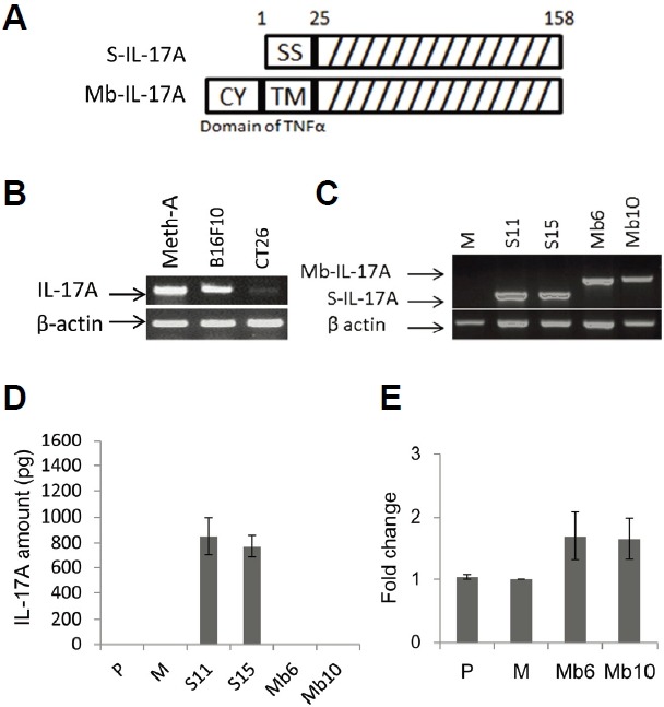
Generation of CT26 clones stably expressing S-IL-17A or Mb-IL-17A. (A) The construct of the secretory form of IL-17A (S-IL-17A) was composed of signal sequence (1–25 a.a.) and IL-17A domain (26–158 a.a.). That of the membrane-bound form (Mb-IL-17A) was composed of the cytoplasmic domain (from −75 to −45, CY), transmembrane fragment (from −44 to −24, TM) and 19 extracellular amino acids (from −23 to −5) of murine TNF-α and the IL-17A domain (26–158 a.a.). (B) Several tumor types were tested for endogenous expression of IL-17A. Meth-A; Meth-A fibrosarcoma, B16F10; B16 melanoma and CT26; CT26 colon cancer. (C) S-IL-17A transfectants (S11, S15) and Mb-IL-17A transfectants (Mb6, Mb10) that strongly expressed IL-17A at the mRNA level, were used in following experiments. M; a mock-vector transfected CT26 clone. (D) IL-17A expression in the culture supernatant of CT26 clones was analyzed by ELISA. Wild-type CT26 cells (P) and a mock-vector transfected CT26 clone (M) were used as negative controls. (E) Membrane-bound IL-17A was stained with polyclonal anti-mouse IL-17A antibody and analyzed by flow cytometry. Each transfectant was also stained with the isotype control and Cy-3-conjugated secondary antibody. Mean florescence intensity was determined by subtracting the basal level from each sample. Data are shown as fold change relative to the mock control (M). Data are means ± SEM of three independent experiments.
