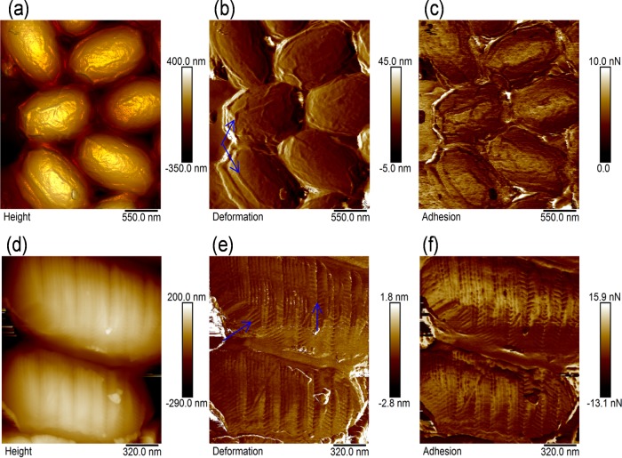
FIG 2.
AFM images of the Bacillus anthracis spore surfaces without (a to c) and with (d to f) the tip-induced surface marks on the mica substrate. The top row includes the height (a), deformation (b), and adhesion (c) image of the intact, native surface of the spores. The bottom row includes the height (d), deformation (e), and adhesion (f) image of the patterned surface of the spores. The images are plane-fitted using the line-by-line algorithm and rescaled for visual clarity.

