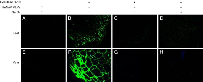
FIG 1.
Binding of HuNoV VLPs to lettuce tissues. Immunofluorescence microscopy was performed on lettuce paraffin slides. NoV/GII.4/HS194 VLPs attached to lettuce green leaf lamina (A–D) and vein (E–H). (A and E) The control sample was not subjected to cellulase R-10 digestion before incubation with NoV VLPs. (B and F) Samples were digested with cellulase R-10 and then incubated with NoV VLPs. (C and G) Samples were digested with cellulase R-10 but not incubated with NoV VLPs. (D and H) Samples were digested with cellulase R-10 and then exposed to NaIO4, followed by incubation with NoV VLPs. The binding signal (green) was detected by primary antiserum against NoV/GII.4/HS194 VLPs and the Alex Fluor 488-conjugated goat anti-guinea pig IgG antibody. Nuclei were stained with 4′,6-diamidino-2-phenylindole (DAPI, blue). All pictures were taken using the same exposure time.

