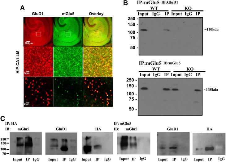
Fig. 1.
mGlu5 and GluD1 colocalize and coimmunoprecipitate in the hippocampus (HIP). (A) Colabeling with GluD1 and mGlu5 was performed in fixed wild-type (WT) hippocampal sections. Confocal imaging demonstrates colocalization of GluD1 (red) and mGlu5 (green) punctas as indicated by yellow colabeling. CA1-LM, stratum lacunosum moleculare field of CA1. (B) Coimmunoprecipitation studies were performed where mGlu5 was immunoprecipitated from hippocamapal synaptosomal membrane fraction preparation, followed by western blotting for GluD1 and mGlu5. GluD1 protein was found to immunoprecipitate with mGlu5 protein. Experiments were repeated five times with protein collected from separate animals, and similar results were obtained. No GluD1 pulldown was observed in GluD1 KO tissue and when IgG alone was used, demonstrating specificity of immunoprecipitation. (C) mGlu5 and GluD1 interaction was tested in HEK293 cells. Cells were transfected with mGlu5 and HA-GluD1, and pulldown of mGlu5 or HA was performed from the protein lysate. GluD1 was found to coimmunoprecipitate with mGlu5. In addition, immunoprecipitation of HA (HA-GluD1) resulted in pulldown of mGlu5. The experiment was repeated five times with similar results. IB, immunoblot.

