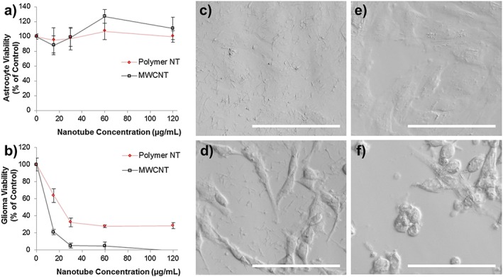
Figure 6.

Cytotoxicity analysis of the polymer nanotubes in comparison with multi‐walled carbon nanotubes (MWCNT) when incubated for 24 h on astrocytes (A) and C6 glioma cells (B) showing that polymer nanotubes exhibited lower toxicity than MWCNT for the C6 glioma, but not for astrocytes where both nanomaterials were tolerated at the concentrations tested (n = 4, error bars represent +/−standard deviation). Light microscopy after 4 h of incubation with the polymer nanotubes (60 µg/mL) shows an even coverage across the well bottom of both astrocytes (C) and C6 glioma cells (D); however after 24 h and three washes with phosphate buffered saline, the astrocytes are largely devoid of nanotubes (E), yet C6 glioma cells have many nanotubes associated with their membrane (multiple thin parallel lines). And many are rounded or washed off the plate completely (F). Scale bars represent 100 µm.
