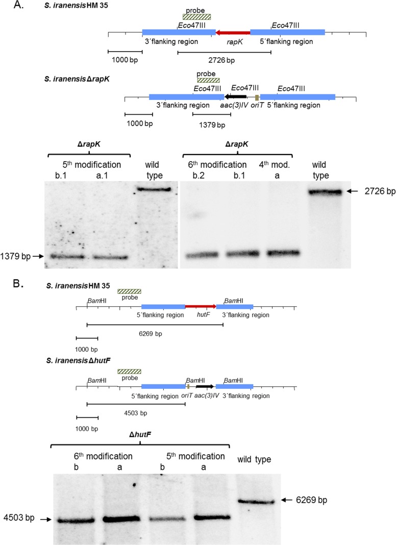
FIG 4.
Southern blot analysis. (A) S. iranensis rapK deletion mutant. The genomic locus was replaced by an apramycin resistance cassette. Genomic DNA was cut with Eco47III. An 801-bp PCR fragment encoding a rapK downstream sequence was used as the probe. ΔrapK deletion mutants (lanes 1, 2, 4 and 6) were characterized by a band of 1,379 bp; the typical 2,726-bp band of the wild type (lane 3 and 7) had disappeared. (B) S. iranensis hutF deletion mutant. The wild-type S. iranensis HM 35 strain was replaced at the locus of hutF by an apramycin resistance cassette. Genomic DNA was digested with BamHI. A 1,047-bp PCR fragment containing the upstream sequence of hutF was used as the probe. In ΔhutF deletion mutants (lanes 1 to 4), the characteristic 6,269-bp band of the wild type (lane 5) disappeared. Instead, a band of 4,503 bp represented successful gene replacement.

