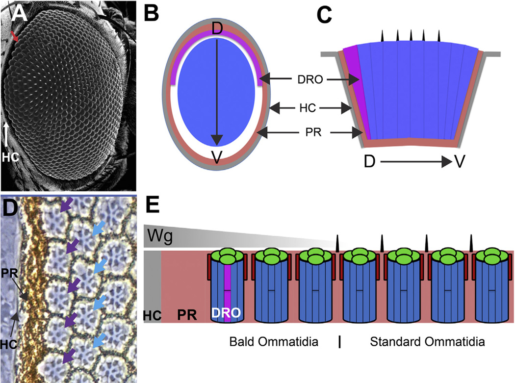
Fig. 1.
Peripheral features of the fly eye. (A) Scanning EM image of adult fly eye. The array of facets and bristles are the most prominent features. The head capsule (HC) circumscribes the eye (white arrow), and the outermost ommatidia lack bristles (red arrow). (B) Schematic cross section through the eye highlighting: the head capsule (HC grey); the pigment rim (PR pink), and DRO (purple). The region of bald ommatidia is white, and the array of standard ommatidia throughout the main body of the eye is blue. (C) Schematic longitudinal section through the eye. (D) Phase contrast image of the dorsal peripheral eye. The head capsule is seen as a grey strip adjacent to the pigment rim. The first row of ommatidia are DRO (purple arrows) indicated by the large R7 rhabdomeres. In the second row, the R7 rhabdomeres are normal (blue arrows). (E) Schematic longitudinal section through the peripheral dorsal eye. The head capsule secretes Wingless (Wg) which diffuses (grey triangle) into the retina and directs the specializations.

