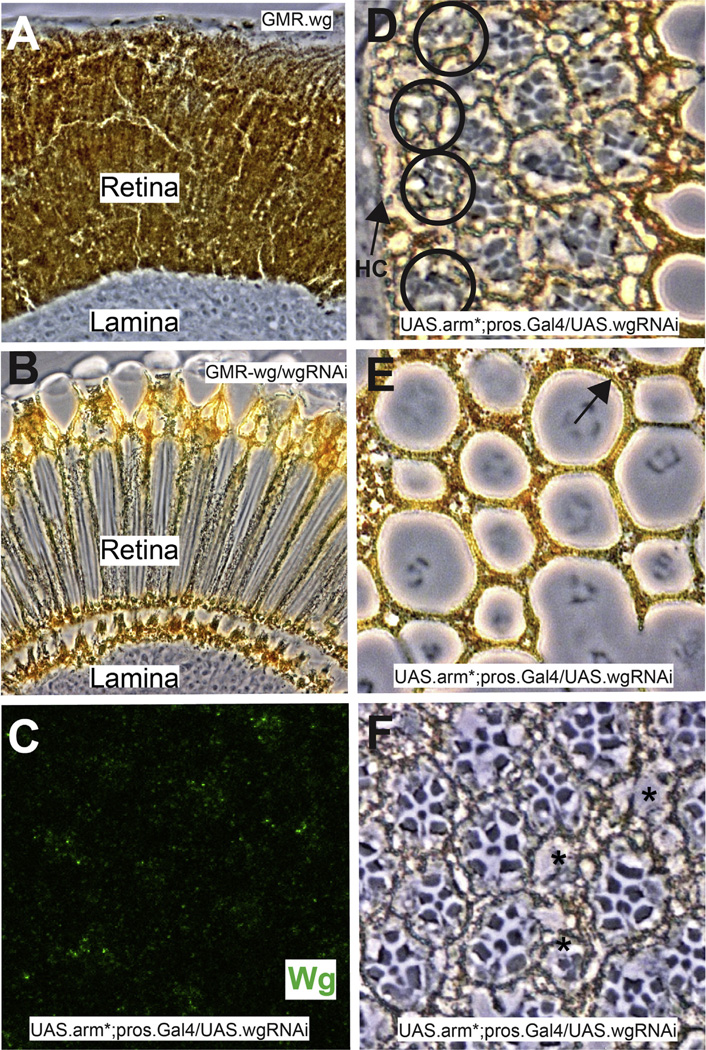
Fig. 4.
The effects of wgRNAi. (A–C) Demonstrations of the efficacy of wg RNAi. (A) Shows a longitudinal section through a GMR.wg eye in which the entire retina appears as pigment rim. (B) When wg RNAi is simultaneously expressed (GMR.Gal4; UAS.wg/UAS.wgRNAi) there is a rescue of the eye back to wild type structure. (C) Image of a Wg-stained (green) 32 h APF pros.Gal4 UAS.arm* UAS.wg-RNAi, showing dramatically reduced Wg expression (compare with Fig. 3C). (D–F) Images of adult pros.Gal4 UAS.arm* UAS.wg-RNAi eyes. (D) The most peripheral ommatidia survive (black circles) bearing disrupted photoreceptor arrays. (E) The lenses are disrupted but 1 °PCs are present (arrow). (F) The photoreceptors array is disrupted, but individual cells appear healthy.

