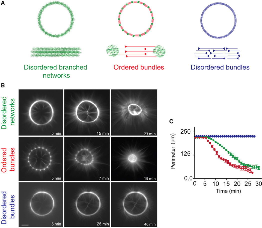
Figure 1. Architecture-Dependent Contractility of Actin Rings.
(A) Schematic representation of the different types of actin architecture: disordered networks, ordered bundles, and disordered bundles, respectively.
(B) Contraction dynamics of the different actin rings, induced by myosin motors. Time is indicated in each picture. Scale bar, 25 µm.
(C) Measured ring perimeter, for each type of ring, as a function of time. The disordered networks (green) and ordered bundles (red) both contract within a few minutes following assembly, whereas disordered bundles (blue) are not contractile within the same time interval. Each curve was obtained by averaging over a dozen of different patterns. Error bars represent SEM. Conditions: 2 µM actin, 6 µM profilin, 100 nM Arp2/3 complex, and 16 nM myosin VI. 300 nM ADF/cofilin was added to the reaction to obtain the ring made of disordered bundles.

