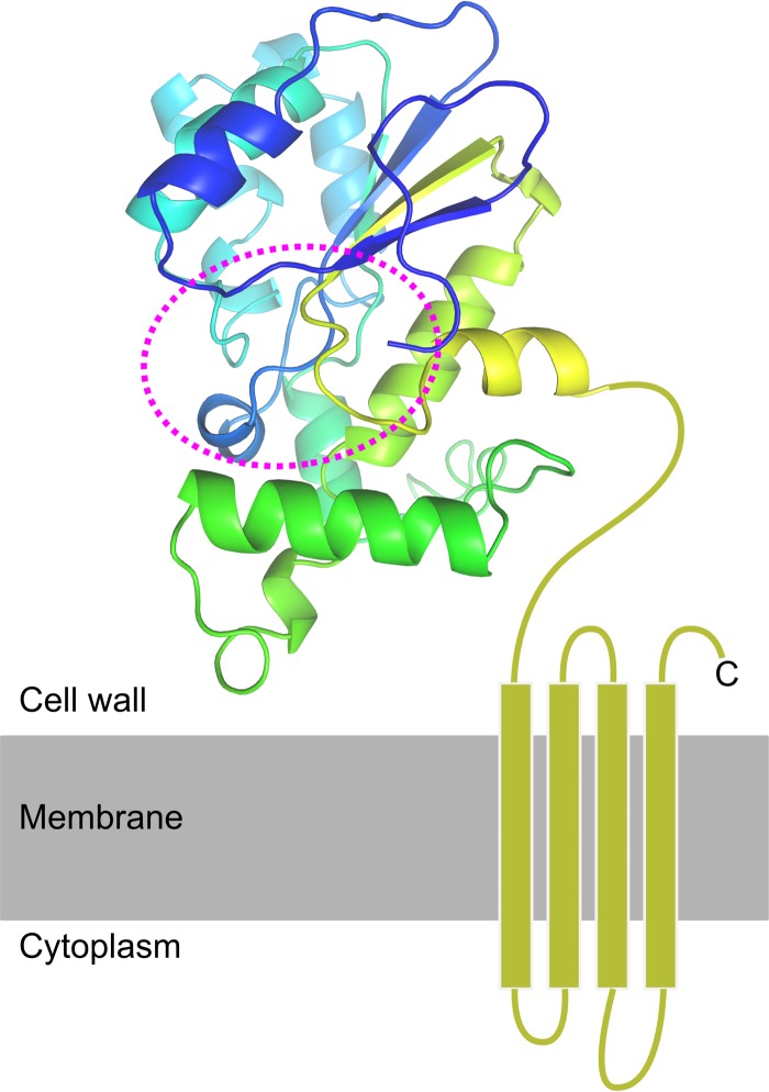
FIG 4.
Predicted structure of PrgY. The extracellular part of PrgY, here shown as a cartoon representation, was modeled using Phyre2 and colored from the N terminus (blue) toward the C-terminal end of the model (yellow). The C-terminal domain, which could not be modeled, is predicted to contain 4 transmembrane helices, shown here as rectangles in a membrane. The predicted active site, based on the homology of PrgY to the Tiki metalloproteases (46), is shown within the dashed line.

