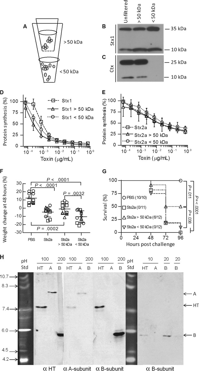
FIG 3.
Stx A and B subunits can dissociate without a loss of activity. (A) Purified holotoxin was separated into fractions of <50 kDa and >50 kDa by use of 0.5-ml concentrating filters until less than 10% of the volume remained, and the fractions above and below the filter were adjusted to the original volume with PBS. (B and C) Partitioning of purified Stx1 (70 kDa; 5 μg ml−1) (B) and cholera toxin (84 kDa; 5 μg ml−1) (C) was examined by Western blotting. (D and E) Protein synthesis inhibition of the unfiltered and filtered fractions of Stx1 (D) and Stx2a (E) was assessed in vitro using Luc2P Vero cells. (F and G) In vivo toxicity of fractionated Stx2a. (F) Mouse weights (circles, trial 1; squares, trial 2; and triangles, trial 3) 48 h after injection. Data are means ± standard deviations. (G) Kaplan-Meier survival curves demonstrating toxicity of Stx2a in all fractions. The data represent the results of three independent trials. Statistical analysis was performed using unpaired Student's t test in GraphPad Prism 5.0. (H) Native gel isoelectric focusing. Purified Stx1 holotoxin (HT) and Stx1 A and B subunits were loaded at 200, 100, 20, or 10 ng, as indicated, into pH 3 to 10 isoelectric focusing gels, followed by characterization by Western blotting. The first three Western blot panels show replicate samples loaded into the same gel, which was cut into strips and probed with a rabbit polyclonal antibody to Stx1 (α HT), a monoclonal antibody to the Stx1 A subunit (α A-subunit), or a monoclonal antibody to the Stx1 B subunit (α B-subunit). The purified A subunit migrated at pH 8.8, and the purified B subunit migrated at pH 5.7. When samples were loaded at 100 ng (140 nM), a single band was seen for the holotoxin, at pH 7.6. When samples were loaded at 10 ng (14 nM), only the B subunit was detected in the holotoxin lane, and it comigrated with the purified B subunit.

