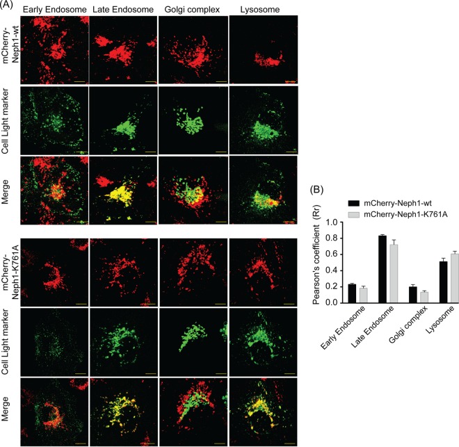
FIG 10.
Loss of Myo1c binding does not influence distribution of Neph1 into various subcellular compartments. To confirm the identity of Neph1-containing vesicles, podocytes stably expressing either mCherry-Neph1-wt or mCherry-Neph1-K761A were colabeled with markers of the early endosome, late endosome, Golgi complex, and lysosome using cell light reagents. Live-cell imaging was performed using confocal microscopy, and deconvoluted images were constructed and are presented (A). Single-plane images were used for analyzing colocalization of Neph1-wt and Neph1-K761A with the early endosome, late endosome, Golgi complex, and lysosome using ImageJ software. (B) Pearson's correlation coefficients are presented as means ± SEM. Scale bars represent 10 μm.

