FIG 4.

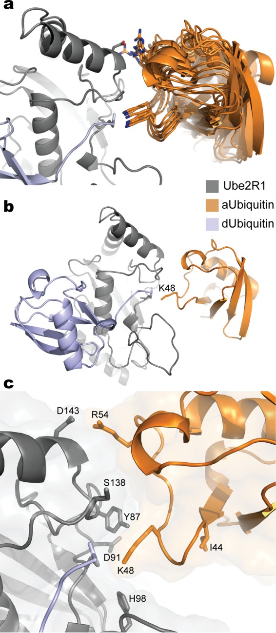
Molecular models of the Ube2R1-donor ubiquitin/acceptor ubiquitin complex after constraining Arg 54 in ubiquitin and Asp 143 in Ube2R1 to be in proximity. (a) Ribbon diagrams for 10 models randomly selected from an ensemble derived from the acceptor ubiquitin refinement procedure. Nitrogen atoms on Arg 54 and Lys 48 are blue; oxygen atoms on Asp 143 are red. (b) Ribbon diagram of a representative model from the ensemble showing the entire Ube2R1-donor ubiquitin/acceptor ubiquitin complex. (c) Closeup view of the Ube2R1-acceptor ubiquitin interface highlighting the locations of key residues that have previously been shown to participate in catalysis. Notice that Ube2R1 residue Asp 91 and acceptor ubiquitin residue Ile 44 are not located at the predicted Ube2R1-ubiquitin interface. Images were generated in Pymol.
