FIG 6.

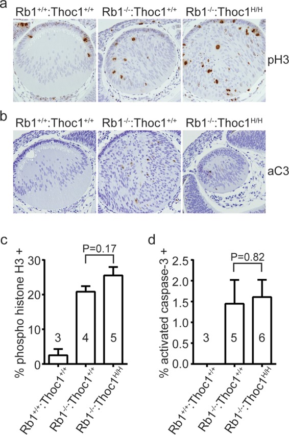
Phenotypes associated with Rb1 loss in the eye lens. (a) Eye sections from E13.5 embryos of the genotypes indicated were immunostained for the cell proliferation marker phosphorylated histone 3 (pH3). Representative sections imaged by bright-field microscopy with a 20× objective are shown. Note the cellular disorganization. (b) Eye sections immunostained with the apoptosis marker activated caspase 3 (aC3). (c) The fraction of cells staining positive for pH3 was measured in E13.5 embryos of the genotypes indicated. The graph shows the mean values and standard errors, and sample sizes are shown in or near the bars. The P value derived with the Student t test by comparing the genotypes indicated is shown. (d) The fraction of cells immunopositive for activated aC3 was measured and graphed as described above.
