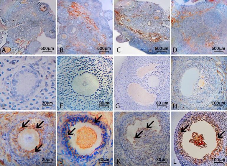
Fig. 2.
Immunohistochemical staining for caspase-3: (A) Vitamin E-received group on day 14, (B) CPM alone-exposed group on day 14, (C) Vitamin E-received group on day 24 and (D) CMP alone-exposed group on day 24 after exposure. Co-administrating of vitamin E significantly reduced biosynthesis of caspase-3 protein on days 14 and 24 after exposure to CPM. Second row is representing intact early secondary (E), late secondary (F), tertiary (G) and graafian (H) follicles, which are not expressing caspase-3. However, the atretic early secondary (I), late secondary (J), tertiary (K) and graafian (L) follicles are representing intensive caspase-3 expression. Arrows are representing chromogens for caspase-3, (IHC

