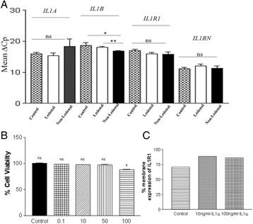
Fig. 8 (abstract P22).

a IL1A, IL1B, IL1R1 and IL1RN transcript levels in skin of vitiligo patients (n = 12) and controls (n = 12). Significant increase of IL1B transcript levels in non lesional skin compared to lesional skin of vitiligo patients and control skin. (p = 0.0021 and p = 0.0290 respectively) was observed. However, no significant difference in the transcript levels of IL1A, IL1R1 and IL1RN was observed. b MTT cell viability assay: IL1-α (100 ng/ml) showed significant decrease in cell viability of NHM at 48 hrs (n = 3, *p < 0.0210). However, at lower concentrations (0.1-50 ng/ml) IL1-α did not show any significant change in the % cell viability. c The NHM were treated with 10 ng/ml IL1α and 100 ng/ml IL1α and subsequently IL1R1 levels were measured by Flow cytometry after 48 hrs of treatment. Results showed significant increase (~22 %) in the membrane expression of IL1R1 upon exogenous IL1-α stimulation to NHM
