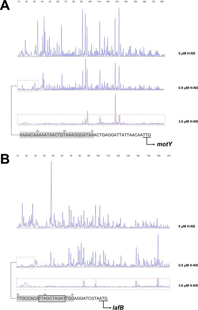
FIG 5.
Identification of the sequence of the H-NS-protected regions of the motY (A) and lafB (B) promoter by DNase I protection footprinting. A concentration of 0.075 μM probe PmotY and PlafB covering the entire promoter region of motY and lafB was incubated with H-NS (at 0.9 μM and 3.6 μM) in the EMSA buffer. The promoter fragments were labeled with 6-carboxyfluorescein (FAM) dye. The regions protected by H-NS from DNase I cleavage are indicated with red dotted boxes. The sequences of the high-affinity H-NS binding region are shown at the bottom (with the protected region in gray shading), and the start codons are underlined. The black box denotes the conserved H-NS binding motif.

