FIG 1 .
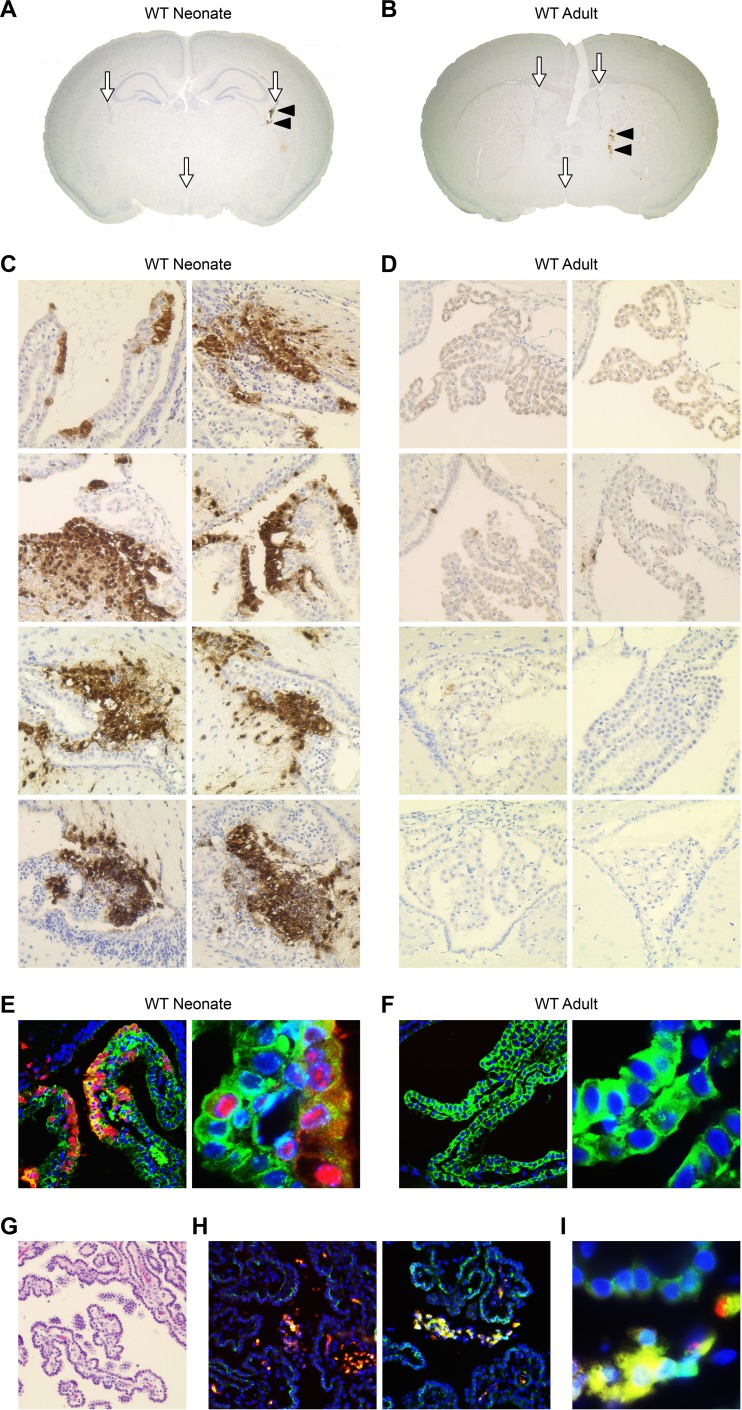
The choroid plexus (CP) is susceptible to HSV-1 early in infection of the newborn brain but not the adult brain. (A) Coronal section of WT newborn murine brain (original magnification, ×20) demonstrating the CSF-filled ventricles which contain the CP (white arrows) and HSV-1-positive cells in the CP, stained brown for HSV antigen (black arrowheads). (B) Coronal section of WT adult murine brain (original magnification, ×20) demonstrating ventricles with CP (white arrows) and HSV infection of brain parenchyma (black arrowheads). (C, D) Representative IHC for HSV antigen (original magnification, ×200) of neonatal WT mice (n = 4) inoculated i.c. with 103 PFU of WT HSV-1 (C) and adult WT mice (n = 4) inoculated i.c. with 104 PFU of WT HSV-1 (D) and perfused at 48 h postinfection. The CP in the newborn brain is positive (stained brown for HSV antigen) in all infected animals (rows) at different HSV-positive foci in the brain (columns). The adult CP is negative for HSV. (E, F) Representative IF of the newborn (E) and adult (F) murine CP for HSV (red), E-cadherin (green), and DAPI (blue) (original magnifications, ×200 [left] and ×1,000 [right]). The newborn mouse brain demonstrates colocalization of HSV and E-cadherin (merge, yellow). The adult murine CP is negative for HSV antigen. (G) Hematoxylin-and-eosin staining of HSV-infected human newborn CP. (H) Dual IF for HSV (red) and E-cadherin (green) in a human newborn case of HSV encephalitis (original magnification, ×200). (I) HSV-infected newborn human CP at higher magnification (×1,000). HSV-positive cells (red) are frequently detected in the CP fibrovascular stroma and in the E-cadherin-positive CP epithelium (merge, yellow).

