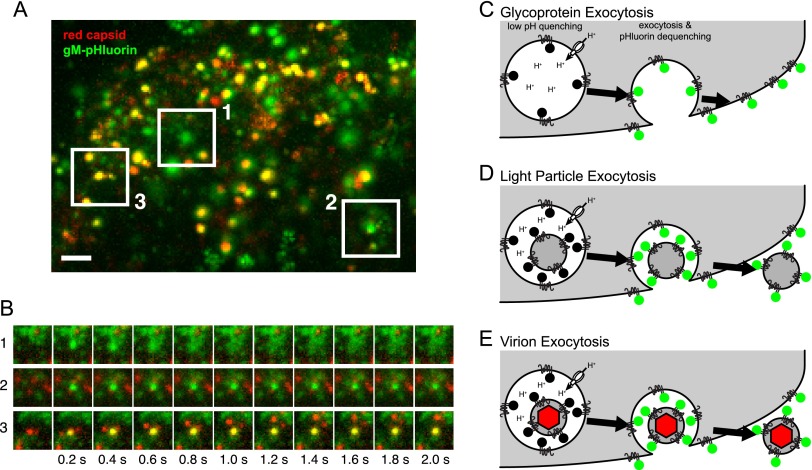
FIG 1 .
Three classes of viral exocytosis. (A) Cells infected with PRV expressing gM-pHluorin (green) and a red capsid tag were imaged beginning at 4.5 h postinfection. Image is a maximum-difference projection, to accentuate areas where gM-pHluorin intensity increases rapidly, projected over a 13-min time course. Boxed areas indicate 3 classes of viral exocytosis events. Scale bar represents 2 µm. (B) Still images corresponding to the boxed areas in panel A. (C to E) Schematics to aid in interpretation of viral exocytosis classes. (C) Schematic of glycoprotein exocytosis, corresponding to box 1 in panel A. gM-pHluorin in a secretory vesicle is quenched in its acidic lumen (black circles). Upon exocytosis, pHluorin is exposed to neutral extracellular medium, becomes fluorescent (green circles), and diffuses into the plasma membrane. (D) Schematic of light-particle exocytosis, corresponding to box 2 in panel A. Upon exocytosis, gM-pHluorin incorporated into light particles becomes fluorescent (green circles) and remains punctate. (E) Schematic of virion exocytosis, corresponding to box 3 in panel A. gM-pHluorin incorporated into a virion is quenched (black circles), but the red capsid tag is not (red hexagon). Upon exocytosis, gM-pHluorin becomes fluorescent (green circles) and remains colocalized with the red capsid.

