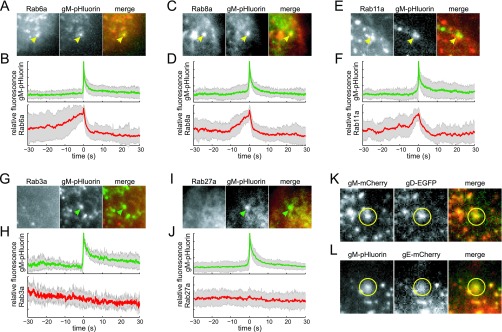
FIG 2 .
Glycoprotein exocytosis. Cells were transduced to express mCherry-tagged Rab proteins, infected with PRV expressing gM-pHluorin, and imaged beginning at 4.5 h after PRV infection. Exocytosis events corresponding to glycoprotein vesicles were selected for analysis (see Fig. 1C). (A and B) Rab6a is associated with gM-pHluorin exocytosis. (C and D) Rab8a is associated with gM-pHluorin exocytosis. (E and F) Rab11a is associated with gM-pHluorin exocytosis. (G and H) Rab3a is not associated with gM-pHluorin exocytosis. (I and J) Rab27a is not associated with gM-pHluorin exocytosis. (A, C, E, G, and I) Still images at the moment of exocytosis show colocalization (yellow arrowheads) or lack of colocalization (green arrowheads) with the indicated proteins. All images are of a 7.2-µm area. (B, D, F, H, and J) Fluorescence divided by fluorescence at time 0 (f/f0) ensemble averages of gM-pHluorin (top, green lines) and indicated Rab protein (bottom, red lines) over a 60-s time course. Shaded areas represent standard deviations. (K) Glycoproteins gM and gD can undergo exocytosis together (yellow circles). (L) Glycoproteins gM and gE can undergo exocytosis together (yellow circles).

