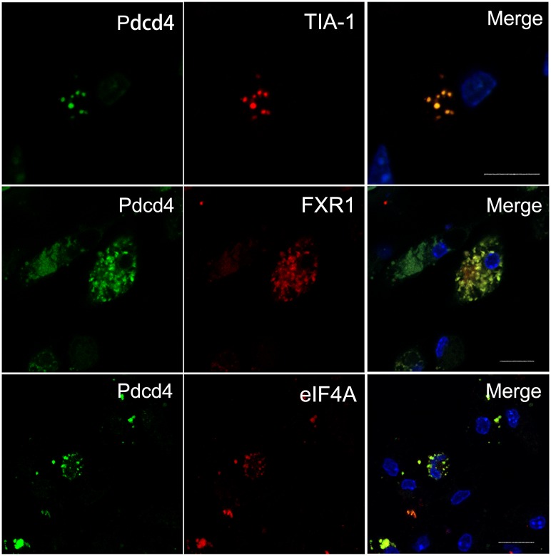
Fig 3. Pdcd4 is co-localized with specific markers of SGs in ox-LDL-treated macrophages.
Primary macrophages from WT mice (n = 6) were stimulated with ox-LDL (50 μg/ml) for 24 h. The expression and co-localization of Pdcd4 and SG markers were examined by immunofluorescence. Pdcd4 immunoreactivity was visualized with FITC (green), TIA-1, FXR1 and eIF4A were detected with rhodamine (red). Cell nuclei were stained with DAPI (blue). The original magnification is 630. Scale bar = 10 μm.

