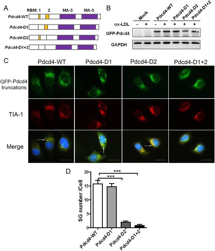
Fig 5. Pdcd4 participates in the SG formation through its RNA-binding region.
(A) Structural models showing the location of two RNA-binding motifs (RBM1 and RBM2) in WT Pdcd4 genes, and three different truncated Pdcd4 genes depleted of RBM1, RBM2, or both, respectively. (B) Representative western-blot for ectopic expression of GFP-Pdcd4 in HepG2 cells transfected with various truncated plasmids. (C) HepG2 cells were transfected with various truncated plasmids and then were stimulated with ox-LDL (50 μg/ml) for 16 h, the SG formation was examined by immunofluorescence. Exogenous Pdcd4 is visualized with GFP (green), TIA-1 was stained with rhodamine (red). Cells were counterstained with DAPI (blue). Arrow head indicates SGs in cells. The original magnification is 1000. Scale bar = 20μm. Data are from three independent experiments. (D) Number of SGs in HepG2 cells transfected with various truncated plasmids. Data are presented as mean ± s.e.m. ***P <0.001.

