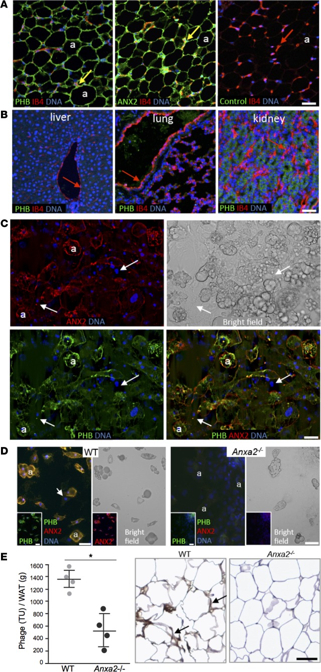
Figure 1. PHB and ANX2 coexpression on WAT endothelium and adipocytes.
(A) Immunofluorescence (IF) on serial mouse white adipose tissue (WAT) paraffin sections with PHB and ANX2 antibodies or a non–immune IgG (control) and green fluorophore–conjugated secondary antibodies. Endothelium (arrows) is counterstained with isolectin B4 (IB4, red). Green/red channel merging indicates expression of PHB and ANX2 in WAT endothelium (yellow). PHB/ANX2 coexpression is also observed in IB4-negative adipocytes (a). (B) IF analysis of mouse paraffin sections with anti-PHB antibodies/IB4 shows that liver, lung, and kidney PHB expression is not detectable in the endothelium (arrows). (C) 3T3-L1 cells after adipogenesis induction were subjected (without permeabilization) to IF with PHB and ANX2 antibodies. Arrows indicate localization of both PHB and ANX2 in differentiated adipocytes (a), for which lipid droplets are shown in the bright field image. Arrows represent nucleated nondifferentiated cells, in which PHB and ANX2 are not detectable. (D) ANX2 requirement for PHB localization to adipocyte surface. WAT-derived cells from of Anxa2+/+ (WT) and Anxa2–/– (ANX2-null) mice adherent to plastic in tissue culture were subjected to PHB (green)/ANX2 (red) IF (without permeabilization). Lipid droplets in differentiated adipocytes (a) are shown in the bright field images. Note the lack of both ANX2 and PHB signal in ANX2-null adipocytes. Arrows indicate cells not differentiated into adipocytes. Nuclei are blue (B–D). (E) Homing of the CKGGRAKDC peptide to WAT is inefficient in the absence of ANX2, as revealed by phage recovery (TU/g of WAT) from visceral WAT extracted from WT and ANX2-null littermates i.v.-injected with 1 × 10¹⁰ of CKGGRAKDC-phage transforming units (6 hour circulation). Shown are mean ± SD from n = 4 mice (2 males and 2 females). *P < 0.05 (Student’s t test, WT vs. ANX2-null). Antiphage HRP immunohistochemistry (brown) in sections of WAT demonstrates phage in WAT vasculature in WT mice (arrows). Hematoxylin counterstaining is blue. Scale bars: 50 μm.

