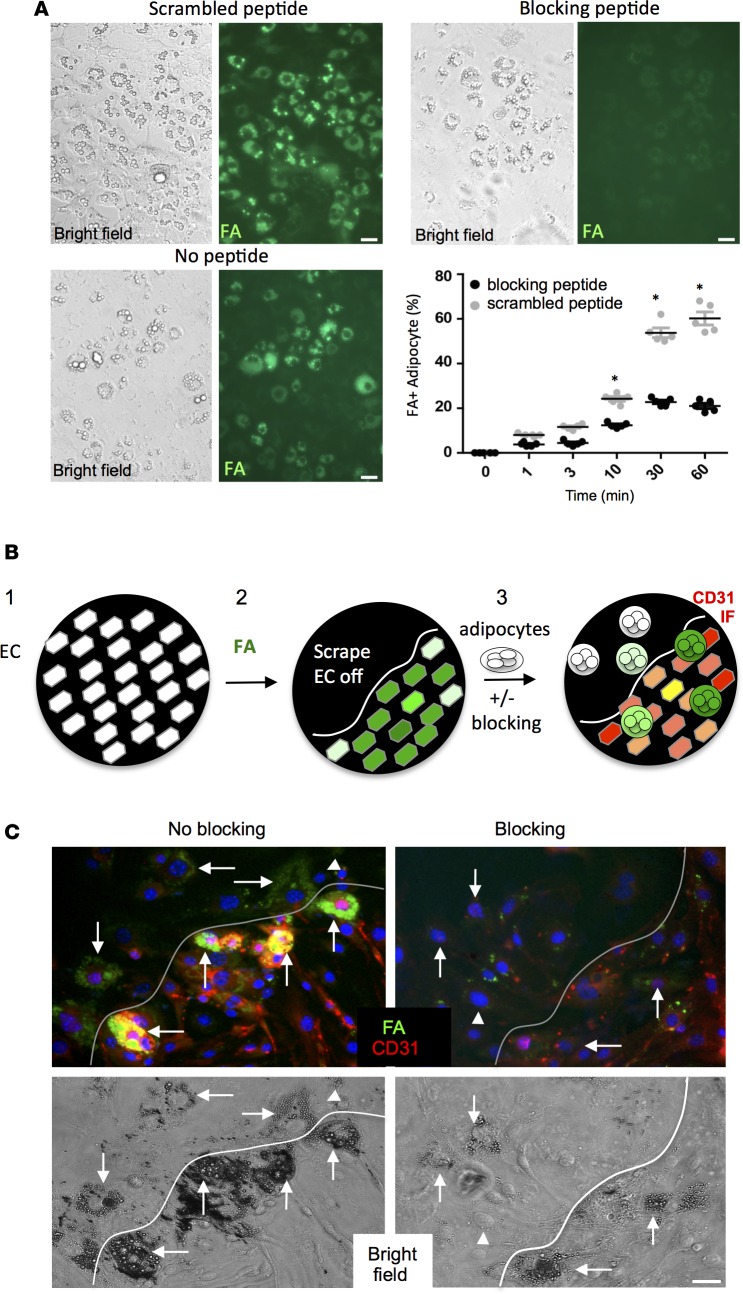
Figure 5. PHB/ANX2 complex mediates lipid transport in WAT.
(A) 3T3-L1 adipocytes were incubated for 30 min. in medium containing 0.1 mM blocking or a scrambled peptide and then treated with 0.3 μM BODIPY-conjugated C16 fatty acid (FA) (green). Note reduced FA uptake upon PHB/ANX2 binding blockade. Graph: data quantified based on the analysis of n = 5 view fields. Error bars ± SEM. *P<0.05 (Student’s t test, blocking vs. scrambled). (B) A schematic of the intercellular transfer assay measuring endothelium-adipocyte FA transfer: (1) bEnd.3 endothelial cells (EC) are plated as a monolayer; (2) EC are incubated with BODIPY-FL-C16 (green fluorescence) fatty acid (FA) and washed, after which a part of the plate is scraped off; and (3) 3T3-L1 adipocytes coated with magnetic nanoparticles are added and forced to the bottom using magnetic field in the presence or absence of the blocking peptide. After washing, CD31 immunofluorescence (red) is performed, and adipocytes that received FA from interacting endothelial cells are observed (green). (C) The intercellular transfer assay performed in the presence of 0.1 mM RDAGRSDALVIYEIGKE scrambled peptide control (no blocking) or AKGRRAEDGSVIDYELI (blocking) peptide. Note that peptide blocking the PHB/ANX2 binding, but not the scramble peptide, prevents FA (green) transfer from the endothelium (red) to adipocytes (arrows). Arrowheads indicate nondifferentiated 3T3-L1 cells. Shown below are bright-field images demonstrating adipocytes attached to plastic and to EC monolayer below the scraped area. Scale bar: 50 μm.

