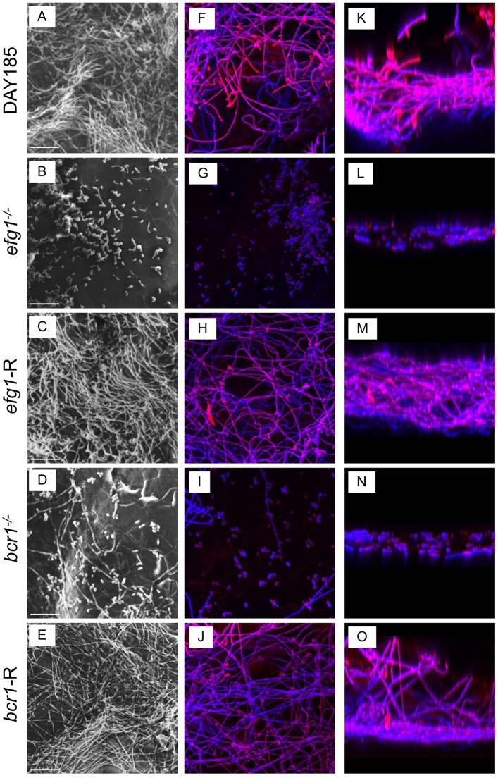
Fig 2. Ex vivo biofilm formation on rat palate tissue.
Excised rat palate tissues were inoculated with 1 x 10⁶ C. albicans DAY185 (A, F, K), efg1-/- (B, G, L), efg1-reconstituted (C, H, M), bcr1-/- (D, I, N) or bcr1-reconstituted (E, J, O) strain and incubated in PBS for 24h at 37°C to allow biofilm growth. The tissues were then processed for SEM (A-E) or stained with calcofluor white (blue; stains fungal chitin in the cell wall) or Concanavalin A-Texas Red conjugate (red; stains mannose in the cell wall and ECM) and examined by fluorescent confocal microscopy to visualize biofilms in XY (F-J) and XYZ (K-O) views. Each panel shows a representative image of 2 repeats. Scale bar = 50 μm.

