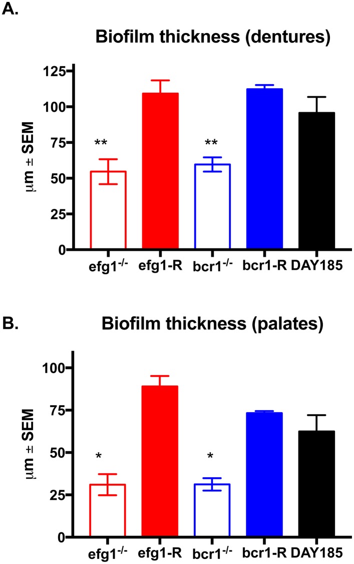
Fig 5. Quantification of biofilm thickness on dentures and palate tissue in vivo.
Equilibrated rats were weaned onto gel diet and fitted with dentures. Rats were given broad-spectrum antibiotics in the drinking water for 4 days prior to inoculation. Rats were inoculated 3x at 3-day intervals with 1 x 10⁹ C. albicans DAY185, efg1-/- or bcr1-/- strain. (A) Dentures and (B) palate tissues were removed from inoculated rats at 4 weeks post-inoculation. Samples were stained with calcofluor white (blue; stains fungal chitin in the cell wall) or Concanavalin A-Texas Red conjugate (red; stains mannose in the cell wall and ECM) and examined by fluorescent confocal microscopy to visualize biofilms. Cross-sectional images of biofilms were visualized by confocal microscopy at 600X magnification, and the depths of biofilms were measured using the Fluoview software. Figure represents cumulative results from 2 independent experiments with 2–3 animals per group and assessment of 5 random areas per animal. Data were analyzed using a one-way ANOVA followed by the Tukey’s post hoc multiple comparison test. *, P < 0.05; **, P < 0.01 compared to the WT control.

