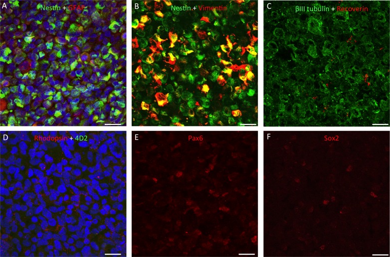
Figure 5.
Immunocytochemistry for retinal markers in sections through a pellet of hRPC, fixed immediately upon cessation of surgery. All cells expressed nestin and βIII tubulin, with some positive staining for GFAP and variable levels of vimentin expression (A–C). Human retinal progenitor cells were found to be largely negative for recoverin (red in [C]), negative for rhodopsin using the 4D2 antibody (green in [D]) but positive for this opsin using a rabbit polyclonal antibody (red in [D]). As shown in adjacent sections, subpopulations of hRPC expressed the retinal progenitor markers Pax6 and Sox2 ([E] and [F], respectively). Proteins of interest are color coded and colocalization appears yellow. Cell nuclei stained blue with DAPI. Scale bars: 20 μm.

