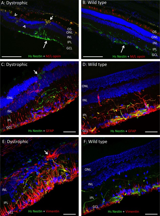
Figure 6.
In the absence of dexamethasone, hRPC survive for 6 months following subretinal injection into RCS dystrophic and wild type rats. Representative images from left eyes showing hRPC stained green with an antibody to human-specific nestin (Hs Nestin) 6 months after grafting into the RCS dystrophic (A, C, E) and WT (B, D, F) retina. Human cells were located in the inner retina where they extend processes into IPL, with human material also seen at the vitreoretinal interface (large arrows in [A] and [B]). Human processes were negative for M/L opsin but positive for GFAP and vimentin to varying degrees (yellow colocalization in [C–F]). Small arrows in (A) and (C) indicate macrophages in the vicinity of the injection site, while arrowheads highlight hRPC processes extending along the outer plexiform layer in (A). Images in (A) and (C) are from rats injected with 200 K hRPC, while all others are from rats injected with 50 K hRPC. Arrow in (E) indicates GFAP-positive subretinal scarring. IPL, inner plexiform layer, outer segments (OS). DAPI staining in blue and proteins of interest color-coded. Scale bars: (A–B) 200 μm, (C–F) 50 μm.

