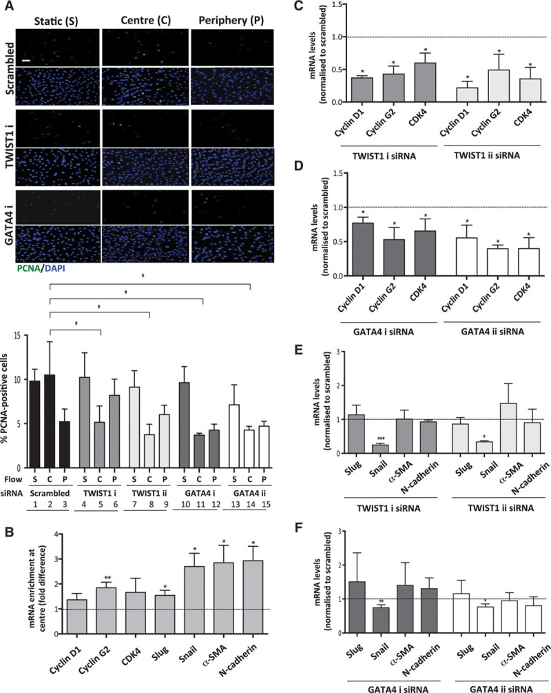
Figure 5.

TWIST1 and GATA4 promoted proliferation in endothelial cells (ECs) exposed to low shear stress. Human umbilical vein endothelial cells (HUVECs) were treated with 2 different small interfering RNAs (siRNAs) targeting TWIST1 or GATA4 (designated i and ii) or with scrambled nontargeting siRNA or remained untransfected. Cells were subsequently cultured in 6-well plates before exposure to orbital flow to generate low (center, C) or high (periphery, P) wall shear stress (WSS) for 72 h. Alternatively, cells were maintained under static (S) conditions. A, Cell proliferation was quantified by immunofluorescent staining using anti-proliferating cell nuclear antigen (PCNA) antibodies and costaining using DAPI (4′,6-diamidino-2-phenylindole). Images are representative of those generated in 3 independent experiments using 1 version of the gene-specific siRNA or scrambled control sequences (bar=50 μm). The percentage of PCNA-positive cells were calculated for multiple fields of view in at least 3 independent experiments, and mean values±SEM are shown. *P<0.05 using a 2-way ANOVA. B–F, The expression of cell cycle regulators and endothelial–mesenchymal transition genes was quantified using qRT-PCR. B, The expression level in cells at the center (low WSS) is presented relative to the expression at the periphery (high WSS; normalized to 1; dotted line). C–F, Transfected cells were exposed to low WSS (center). The expression level in cells transfected with gene-targeting siRNA is presented relative to the expression in cells transfected with scrambled control siRNA (normalized to 1; dotted line). Data were pooled from 3 independent experiments, and mean values±SEM are shown. *P<0.05, **P<0.01, and ***P<0.001 using an unpaired t test.
