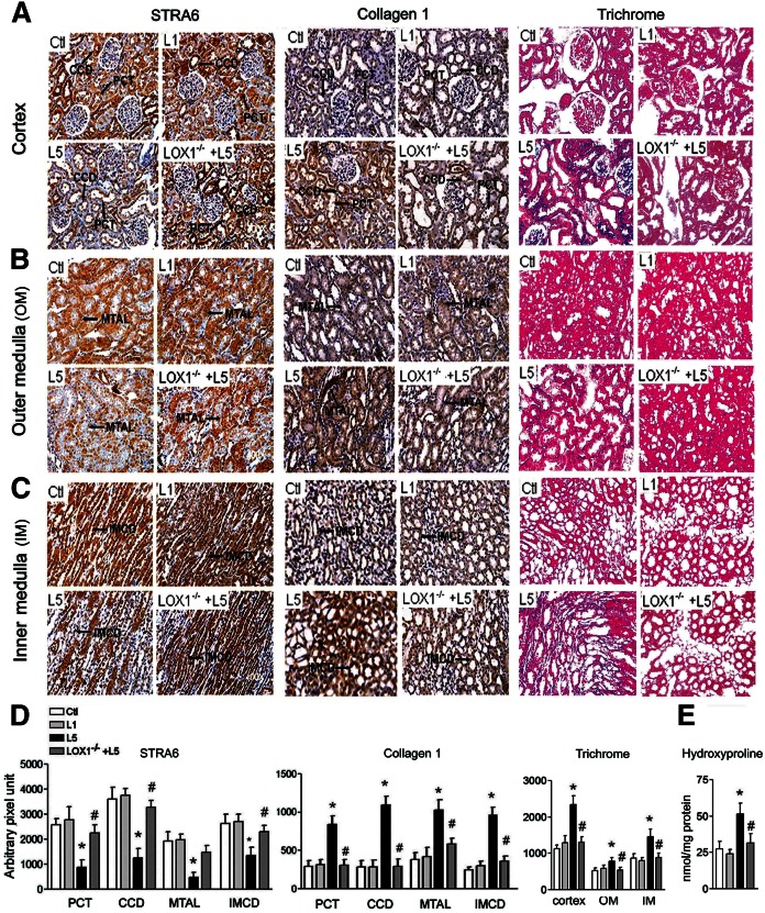
Fig. 2.
L5 decreases STRA6, but increases collagen 1, immunohistochemical staining, trichrome staining, and hydroxyproline concentration in the kidneys. Representative images show immunoreactive staining for STRA6, collagen 1, and Mason’s trichrome stain in cortex (A), outer medulla (B), and inner medulla (C) from saline-injected (Ctl), L1-injected (L1), L5-injected (L5), and L5-injected LOX1−/− mice (LOX1−/−+L5). D: Quantitative analysis showed that STRA6 immunostaining intensity increased but collagen 1 intensity decreased in the PCT, CCD, MTAL, and IMCD in L5-injected mice, as compared with Ctl and L1 mice or L5-injected LOX1−/− mice. Intensity of trichrome stain increased in the cortex, outer medulla (OM), and inner medulla (IM) of L5-injected mice. E: Bar graph shows that hydroxyproline concentration increased in the kidneys of L5-injected mice. All results are represented as mean ± SE. *P < 0.05 versus Ctl and L1; #P < 0.05 versus L5.

