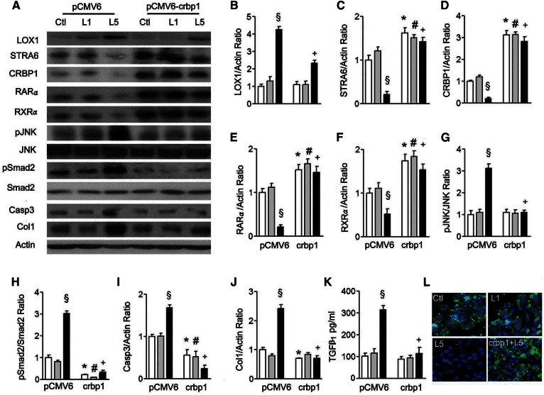
Fig. 5.
The crbp1 gene transfection reverses L5 effects on STRA6 cascades and renal cell injury. A: Western blots show LOX1, STRA6, CRBP1, RARα, RXRα, pJNK, pSmad2, and collagen 1 (Col1) expression in cell lysate of pCMV6-transfected and pCMV6-crbp1-transfected HK-2 cells under PBS (Ctl), L1, or L5 treatment for 24 h (n = 3). Quantitative analysis showed a significant difference for the increase of LOX1/actin (B); the decrease of STRA6/actin (C), CRBP1/actin (D), RARα/actin (E), and RXRα/actin (F); the increase of pJNK/JNK (G), pSmad2/Smad2 (H), caspase 3/actin (I), and collagen 1/actin (J) under L5 stimulation in pCMV6-transfected cells. These changes caused by L5 treatment were reversed in pCMV6-crbp1-transfected cells. K: ELISA showed that pCMV6-crbp1-transfection reversed the elevation of TGFβ1 concentration under L5 treatment in the culture medium of HK-2 cells. L: Cytochemistry images show immunoreactive staining of STRA6 (green fluorescence) was recovered by pCMV6-crbp1-transfection in L5-treated HK-2 cells (crbp1+L5). All results are represented as mean ± SE. §P < 0.05 versus Ctl- and L1-treated pCMV6 groups; *P < 0.05 versus Ctl-treated pCMV6 group; #P < 0.05 versus L1-treated pCMV6 group; +P < 0.05 versus L5-treated pCMV6 group.

